Musculoskeletal Ultrasound
Chapter 14
Chapter 14: Musculoskeletal Ultrasound
435
14
1. History and Background
Musculoskeletal (MSK) ultrasound encompasses a variety of ap-
plications of ultrasound to the appendicular skeleton. The history
of the MSK ultrasound is elegantly described by D. Kane and W.
Grassi in Rheumatology.
5
Briefly, the first published article was by
Dussik KT, Fritch DJ, Kyriazidou, M, et al. in 1958
3
describing the
sonographic signatures of several of the commonly encountered tis-
sues in musculoskeletal imaging. Bruno Fornage, MD, and Marnix
von Holsbeeck, MD, both prominent radiologists, pioneered the
initial advancement of the field of MSK in the 80’s; however with
the introduction of MRI in the early 70’s,ultrasound began receiving
less attention within the United States. A prodigious body of knowl-
edge and research followed and MRI musculoskeletal applications
improved dramatically with field strengths up to 3 Tesla (T) and the
design of joint specific imaging coils, as well as MR arthrography.
Although peer reviewed clinical and research journals provided an
incredible body of evidence supporting the value of musculoskeletal
MRI imaging, artifacts such as “magical angle”
7,12
and “star” effect
on metal implants
9
became well known as limitations to muscu-
loskeletal MRI. Computerized axial tomography (CAT), positron
emission tomography (PET), and computed tomography (CT) are
additional imaging techniques that provide unique and important
diagnostic information,especially with 3D reconstruction in surgical
planning. Plain film radiography remains the diagnostic standard
in any initial joint evaluation.
Early pioneers such as von Holsbeeck,Erickson,Fornage,Martinoli,
Bianchi
15
and more recently Klauser
9
and others saw the benefit of
ultrasound in the evaluation of themusculoskeletal system.With the
advent of high-resolution compact ultrasound units at competitive
prices, and because of the advantage to instantly evaluate targeted
anatomy while performing dynamic maneuvers, ultrasound has
become an attractive extension of the clinical exam.
2. Structure
The discussion of MSK will be subdivided into four (4) primary
categories:
1) ultrasound system parameters
2) tissue imaging characteristics
3) normal and pathologic tissue signatures
4) artifacts
3. Ultrasound System Parameters
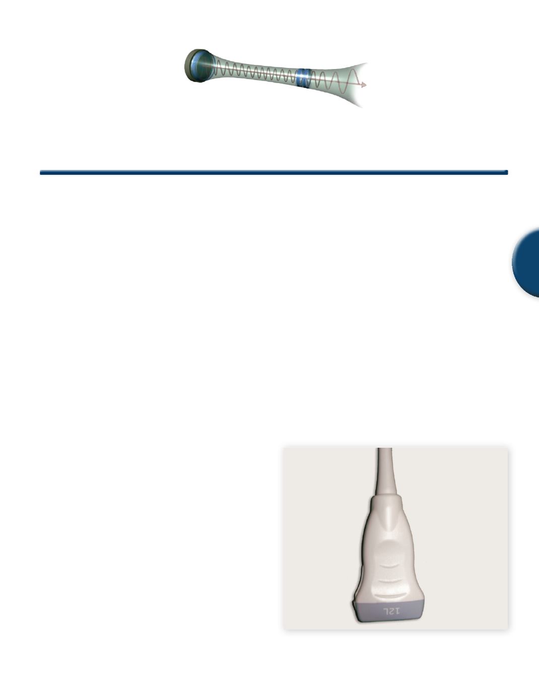
3.1 Linear Array Transducers and Operating Frequency
For optimal MSK imaging, the most important system parameter is
the transducer’s operating frequency range (as indicated in Chapter
6 for imaging in general). The ideal operating frequency yields
adequate penetration to view the desired structures while simultane-
ously providing the resolution necessary to visualize macroscopic
changes and structure variations. Linear and curved linear array
transducers are typically used (See
Figures 1a and 1b
). Typical linear
array operating frequencies are in the 9-20 MHz range.
Fig. 1a
Linear array transducer
Patrick R Meyers BS, RDMS, RDCS RVT


