426
Level 2
Board Level
KEY CONCEPT
The Spectral Window
Presence in Healthy Vessel
Figure 37
displays the Doppler spectrum for blood flow in a normal
common carotid artery. For this spectrum, the sample volume was
relatively small (1.5 mm) and was placed in the center stream of the
flow. Since the flow is laminar,it accelerates and decelerates without
creating eddies or swirling flow patterns. Since the sample volume
is centrally placed in a large vessel where the frictional and viscous
effects are least pronounced, the highest velocities in the vessel are
recorded, of course varying with the changing pressure throughout
the cardiac cycle. By its central placement in a large vessel, the
sample volume does not include the low and zero velocity flow along
the walls of the vessel. All of the velocities recorded in systole are
relatively high, creating a region of no signal at the lower velocities
closer to the baseband. This region of signal absence is called the
spectral window. The spectral window is indicated by the white
arrows in
Figure 37
.
Fig. 37
Spectral window
Absence in Diseased Vessel
As previously discussed, in the presence of turbulence, the velocity
varies significantly, resulting in an entire range of frequency shifts.
Unlike the laminar example we have just discussed, in addition to
the narrowband of frequency shifts about the peak velocity, there is
an entire range of frequency shifts continuing all the way down to
the baseline, and depending on angle, potentially even extending in
the opposite direction. As a result,when performing Doppler in the
presence of turbulence, the spectral window is not present.
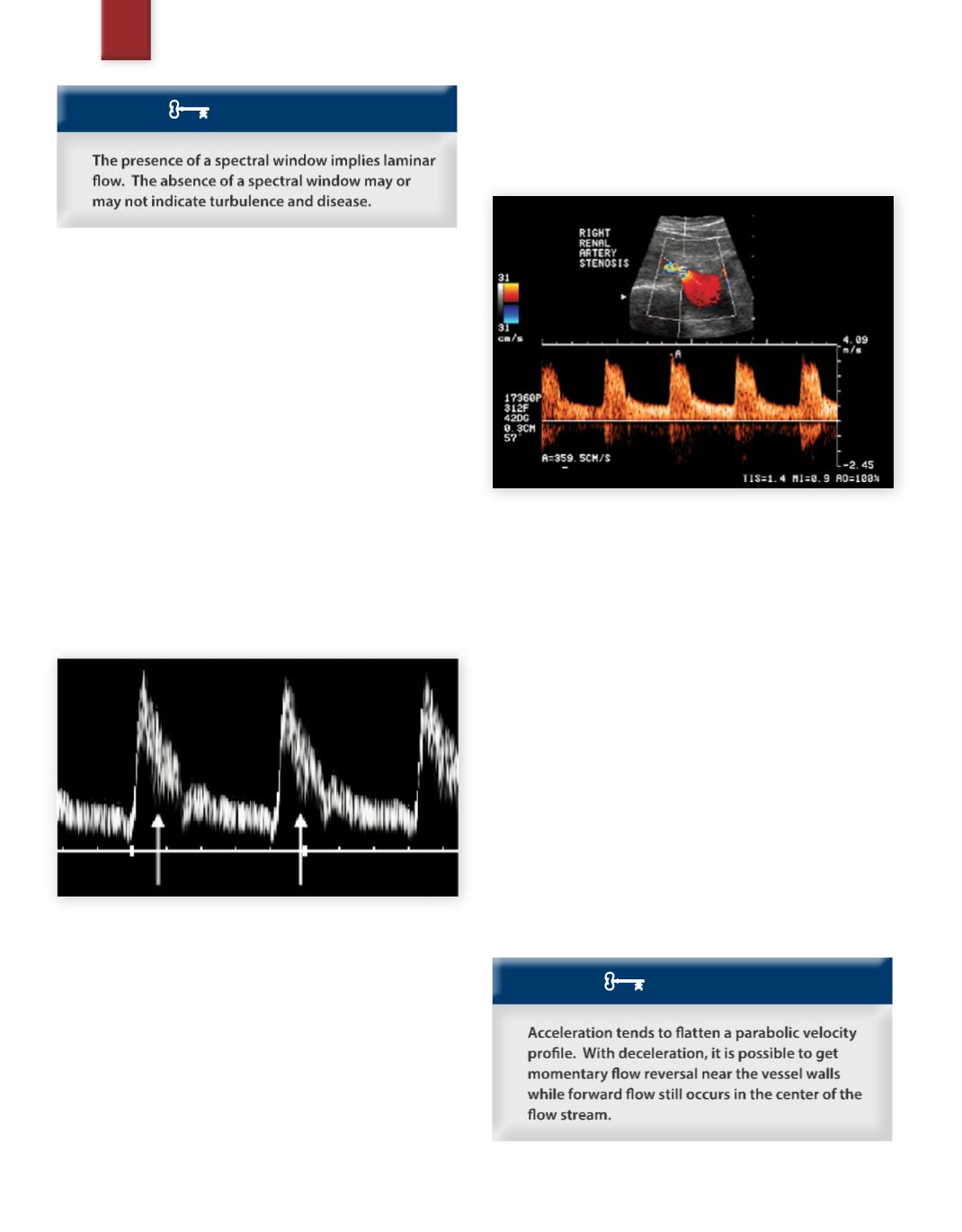
Figure 38
demonstrates the turbulence that occurred in a right renal
artery stenosis. Note that there is no spectral window in systole,
and there is a component of flow in the reverse direction (below
the baseline).
Fig. 38
Spectral display of turbulence
Absence of the Spectral Window
in a Healthy Vessel
There are many reasons which can cause the absence of a spectral
window besides flow disturbance, including:
• CW Doppler (instead of PW Doppler)
• A large PW sample volume (gate size) relative to the vessel
size (small vessels rarely display a spectral window)
• The sample volume too close to the vessel wall (including in the
elevation direction)
• Overgaining of the spectrum
• Flow angle close to 90 degrees
• Spectral broadening artifact
From this list, it is obvious that the absence of the spectral window
does not necessarily imply a hemodynamic state of turbulence.
Therefore, caution must be used when interpreting based on the
absence of the spectral window. Conversely,when a spectral window
does exist, it is evident that there is laminar flow.
KEY CONCEPT
SAMPLE PAGE


