Chapter 15: Focused Ultrasound
455
15
4.2 Other Indications for Focused Ultrasound Therapy
In addition to the three indications listed above, FUS is in various
stages of clinical research for treatment of the following indications:
• Bone metastases
• Low Back Pain Caused by Facet Arthritis
• Osteoid Osteoma
• Breast Cancer and Breast Fibroadenoma
• Refractory Glaucoma
• Benign Thyroid and Primary Hyperparathyroidism
• Multiple Brain Indications
As with the previous indications, more complete discussions are
found in the online image library.
VIEW ONLINE ANIMATION AND IMAGE LIBRARY
4.3 Multiple Brain Indications
Of all of the indications and organs listed,FUS brain procedures may
provide the most promise for this technology. Noninvasiveness,lack
of ionizing radiation,and high precision coupledwith tight focus and
sharp ablation margins are ideal for brain surgery procedures. In
addition, FUS has the potential to noninvasively stimulate or block
brain tissue activity,facilitate a reversible opening of the blood-brain
barrier, and liquefy blood clots. The bony skull, however, provides a
significant acoustic barrier by reflecting, absorbing, and refracting
(and thereby defocusing) the ultrasonic energy directed at the brain.
The varying skull thickness, along with its heterogeneous internal
structure, affects the wave propagation so much that the focal point
is degraded (See Section 5.2 of Chapter 8 on beam aberration).
To overcome these technical difficulties, the solution currently
employed is to use a high power, large aperture (
)
helmet-shaped transducer coupled with skull information derived
from high precision computed tomography (CT) data that is regis-
tered to treatment-day MRI images. The transducer in the helmet
is comprised of many elements, for which the phase can be varied.
By using the three-dimensional CT data and ray acoustic methods,
the therapy systemcalculates the expected phase shifts andmodifies
the phase delays of relevant elements in such a way that the skull
actually refocuses the corrected acoustic wave. To avoid skull heating
and damage to skin and adjacent brain tissue, the systemuses active
degassed water cooling for the skin and a calculated energy density
threshold to avoid thermal damage to the brain cortex. Research-
ers have already used this method, sometimes called transcranial
MRI-guided focused ultrasound (tcMRgFUS), with encouraging
results to treat approximately 50 patients under multiple research
protocols for essential tremor, neuropathic pain, Parkinson disease,
and (limited) tumor ablation.
Using the mechanical effects of FUS on brain tissue is in the early
stages of development. Preclinical work has shown that it is possible
to either stimulate or inhibit brain tissue using pulsating low-level
acoustic energy. Once mature, the potential application of this
method could stop an epileptic seizure or provide high-resolution,
noninvasive deep brain stimulation. Researchers also achieved a
reversible opening of the blood-brain barrier in vivo using a com-
bination of tcMRgFUS and injected ultrasound contrast material.
Noninvasive liquefaction of blood clots was tested as well—ex vivo,
in vivo, and in cadavers, and was shown to be feasible. The near
future is sure to see different approaches for FUS application in the
brain as well asmaturation of the ideas currently under investigation.
5. Parameters of Technology Adoption
Multiple vendors aremanufacturing FUS systems for various clinical
indications,and each is at various stages of regulatory approval in the
different geographical regions. For a review of current applications
view the Online Library.
VIEW ONLINE ANIMATION AND IMAGE LIBRARY
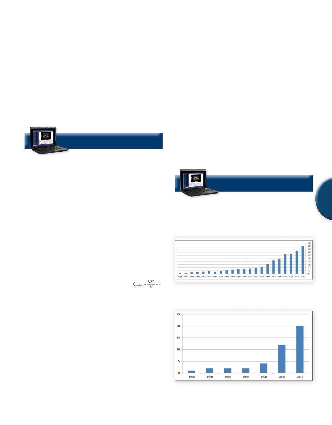
Recently, significant publication and clinical research progress has
beenmade in this area in terms of new indications (
Figures 2 and 3
).
Fig. 2
The number of scientific publications on the subject of
focused ultrasound per year. Source: Web of Science.
(Graph
courtesy of Focused Ultrasound Surgery Foundation.)
Fig. 3
Number of clinical stage indications for focused ultra-
sound reported by year.
(Graph courtesy of Focused Ultrasound
Surgery Foundation.)
SAMPLE PAGE


