Chapter 18: Speckle Tracking and Cardiac Strain
483
18
and frequency. As ultrasound energy passes through tissue, con-
structive and destructive interference (as discussed in Chapter 1)
creates speckle patterns that can be seen within the more familiar
specular reflection of large objects. When imaging the heart,much
of the interference occurs frommyocardial fibers and, even though
the ultrasound image pattern appears “random”, the appearance of
the pattern is periodic and dependent on
1. the tissue properties and tissue architecture
2. the way the ultrasound system“processes” the signal
known as its “transfer function”.
Therefore,the speckle patterns of the exact same portion of myocar-
diummay be different on different imaging systems and with differ-
ent imaging settings, but it looks the same on the same machine at
the same point of the cardiac cycle. The fact that the speckle patterns
are different doesn’t matter so long as they allow precise measure-
ment of heart motion, what we will call “myocardial deformation”.
For decades, ultrasound engineers have searched for methods to
suppress speckle patterns by using various averaging or filtering
methods. This was done in the hopes of “cleaning up”the image and
making it more visually appealing. However, although the speckle
pattern is not related to anatomical structures, there is information
encoded within the speckle pattern. As noted above, speckle pat-
terns are influenced by myocardial tissue and the deformation of
tissue changes the speckle pattern. Therefore, changes in speckle
pattern can be related to deformation of tissue. Ultimately, we can
use the changes in speckle pattern to track moving myocardium
and, even better, to quantify the tissue deformation throughout the
cardiac cycle.
3.2 How Speckle Tracking Works
The most straightforward approach to understand speckle tracking
is to realize that the goal is to track and quantify moving targets in
tissue. An easy analogy is to think of air traffic controllers tracking
moving aircraft targets on their radar screens. Ideally, we would
like to track the path of each plane and not confuse one plane with
another plane that is either nearby or flying in a similar path. Radar
is successful in identifying each plane since each plane has a unique
radio signature (from its transponder and profile).It is important to
note that for the radar to function well, the frame rate of the radar
screen has to be high enough to sample each plane’s motion or their
precise location will not be known and airplanes could potentially
collide. You could imagine a case in which if the radar is too slow,
an aircraft could pass through the controller’s territory without the
controller even knowing the plane was there. In essence, the radar
system must have adequate temporal resolution to be effective.
Similarly,when tracking tissue, temporal resolution is important or
we will not be able to accurately track tissue motion.
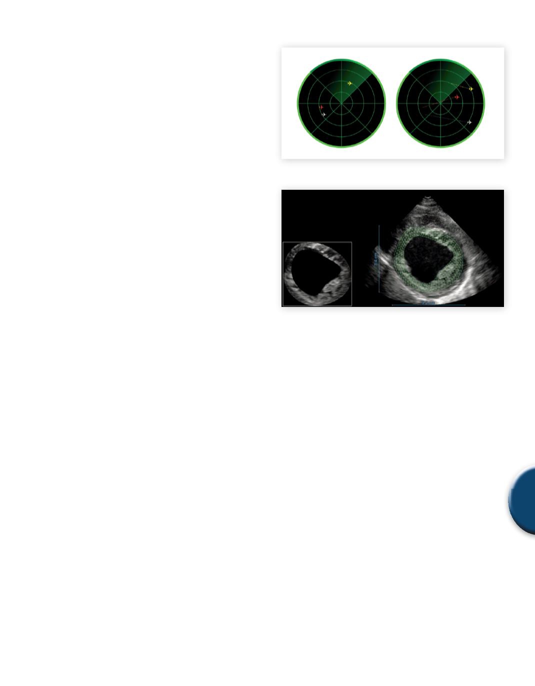
Fig. 6
Radar analogy of how speckle tracking works
Fig. 7
Regional speckle tracking
Motion of tissue contains physiologic information. When ultrasound
is used to examine tissue stiffness of non-cardiac tissues such as
breast or liver, some external force is needed to compress the tissue
resulting in deformation and hence strain. The extent to which
the tissue deforms, its “strain”, is determined by the inherent tissue
properties (e.g. its “stiffness”) and how hard the “push” was (i.e. the
stress.) For the healthy heart,motion occurs withmuscular contrac-
tion without any external forces, or what is referred to as intrinsic
motion. Myocardial motion due to contracting muscle creates its
own deformation, or “strain”. Therefore, on one level, measuring
strain in cardiac settings is simpler in that the tissue deformation
being measured is inherent to the cardiac tissue.
Speckle tracking, like tissue Doppler, is another tool to measure
strain. Unlike tissue Doppler - speckle tracking is not angle depen-
dent. Also, speckle tracking uses fundamental B-mode imaging
which uses one cycle transmit pulses. Essentially, tissue patterns
can be identified, tracked frame-to-frame, and quantified to yield a
measure of strain. Systems use some form of cross-correlation (or
equivalent) to track pixels from frame to frame in non RF speckle
tracking. This is the case for most commercially available systems
measuring cardiac strain. B-mode images are generated in an ul-
trasound system by measuring the amplitude of the envelope of the
acoustic signal without using the phase information. As long as the
systemhas adequate resolution,motion can be tracked anywhere in
the frame. Moreover, this technique is insensitive to overall cardiac
translation (e.g.“swinging in the chest”) since only the deformation
is being tracked.
SAMPLE PAGE


