Chapter 17: IMT Ultrasound Imaging
471
17
Finally, the angle of incidence for acquisition must be determined.
As atherosclerosis often presents asymmetrically, and since the
beam thickness is much smaller than the diameter of the artery,
it is important to longitudinally intersect every analyzed segment
of the arterial cylinder in multiple planes (as shown in
Figure 4
).
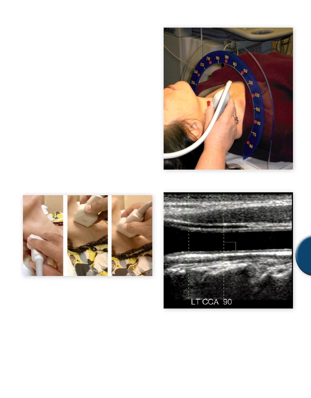
The precision and reproducibility of the angles of incidence can
be enhanced by using a Meijer’s Arc (protractor) to guide probe
placement on the neck (see
Figure 5
). Developed by Rudy Mei-
jer, University Medical Center Utrecht, The Netherlands, the Arc
was designed to standardize single and multi-angle protocols. Of
paramount importance in acquiring images for IMT measurement
is the technologist’s attention to orienting the arterial segment of
interest perfectly parallel to the transducer face, or in other words,
at 90° to the axis of the sound beam (equivalent to 0° incidence).
Recall that specular reflection is very angle dependent. By achieving
a 0° incident angle (see
Figure 6
), the specular reflections from the
blood-intima andmedia-adventitia borders are optimally captured,
eliminating any chance of refraction at those surfaces which would
make for a much less distinct (and less traceable) IMT (see
Figure
7
). This said, the carotid anatomy beyond the CCA becomes less
predictably straight and is often steeply angled relative to the skin
surface. Therefore,CIMTmeasurements in the bifurcation and ICA
regions can be correspondinglymore difficult to accuratelymeasure
and reproduce.
A
B
C
Fig. 4
Analyzing multiple planes of the carotid artery.
A: posterior, B: anterior, C: lateral
Fig. 5
Meijer’s Arc (image courtesy of Rudy Meijer, Meijer Medical
Ultrasound)
Fig. 6
Normal incidence
SAMPLE PAGE


